Advanced Diagnostic Testing
At Village Eyecare we believe our patients deserve the best, so we choose to invest in the latest state of the art equipment. Better technology allows us to practice at the highest level and detect and treat everything from refractive errors to eye diseases sooner and more efficiently. Advanced technologies we offer include: Corneal Topographer, Dry Eye Analyzer, Visual Field Machine, OCT (Optical Coherence Tomography), Anterior Segment Camera, Retinal Fundus Camera, Digital Refraction Systems, Electronic Medical Records, and Patient Education Videos.
Corneal Topography
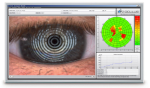
Corneal Topography is a non-invasive imaging technique that creates a three-dimensional map of the surface curvature of the cornea, the outer structure of the eye. Uses of corneal topography include:
- Cornea Disease; the ability to detect irregular conditions invisible to most conventional testing
- Contact Lenses; assisting in fitting contact lenses to optimally fit the cornea
- Refractive Surgery; the corneal topography map is used in conjunction with other tests to determine exactly how much corneal tissue will be removed to correct vision
OCT (Optical Coherence Tomography)
Optical Coherence Tomography, or ‘OCT’ is a powerful new test that can help your doctor identify and manage vision threatening conditions early.
- Scans the structures in the back of the eye
- Generates a highly-detailed, cross-sectional image of the back of the eye
- Detect changes in the eye, caused by diabetes and macular degeneration
- Help doctors quantify abnormal thickening of the center of the retina, known as macular edema
- Help identify subtle, less common retinal conditions such as macular holes
- Monitor changes to the back of the eye, as a result of drug or laser therapy
The OCT procedure is brief, painless, and completely safe. Let the OCT test provide you and your doctor valuable information today that can help preserve your vision for tomorrow.
Retina Imaging
Retina Imaging uses a high-magnification camera connected to a biomicroscope to capture high resolution images of the retina, macula and optic disc. Retina Imaging allows for monitoring the progression of the following conditions:
- Diabetes
- Macular Degeneration
- Glaucoma
- Hypertension
- Choroidal Nevus

Visual Field Test
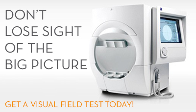
Visual Field Test is an analysis of the peripheral (‘side’) and central vision. Some conditions that cause loss in the Visual Field include:
- Glaucoma
- Diabetes
- Retinal Detachment
- Macular Degeneration
- Multiple Sclerosis
- Strokes
- Brain Tumors

Dry Eye Analyzer
- Tear Quality: the non-invasive tear film break-up time (NIKBUT) measures tear film stability and how long a patient’s tears last. The NIKBUT is automatically measured within seconds.
- Oil Gland Analysis: easily and efficiently images the health of the meibomian glands which are responsible for the lipid (oil) layer of the tear film. The dysfunction of meibomian glands is the most frequent cause of dry eye. Morphological changes in the gland tissue are made visible using the Meibo-Scan.
- Redness Analysis: for the first time it is possible to objectively classigy redness of the eyes completely and automatically using the R-Scan.

“No Puff” Eye Pressures
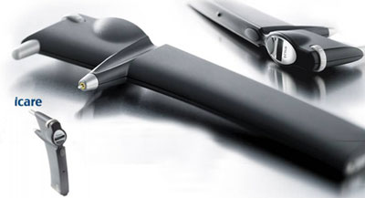
Say good-bye to the dreaded air puff test. Although harmless, many people have a strong dislike of the test and we are pleased to announce we now use Icare tonometers. Icare tonometers are based on unique, patented rebound technology, in which a very light and small probe is used to make a momentary contact with the cornea.
- Quick and painless
- Barely noticeable to patients
- More accurate measurements
- Glaucoma detection & prevention


